
Dental Imaging
OPG / Cephalometric X-Ray
OPG and cephalometric X-rays are specialised dental X-rays used to provide an overview of the teeth, jaws and surrounding facial structures. They are commonly requested by dentists, orthodontists and oral surgeons to assist with treatment planning.
An OPG provides a panoramic image of the upper and lower jaws and teeth, while a cephalometric X-ray provides a side-view image of the skull and facial bones.
Available At
What does an OPG or cephalometric X-ray examine?
An OPG can show the teeth, upper and lower jaws, jaw joints and surrounding structures in a single panoramic image.
A cephalometric X-ray shows the relationship between the teeth, jaws and facial bones and is particularly useful for orthodontic assessment and treatment planning.
Why might I need an OPG or cephalometric X-ray?
Your dentist, orthodontist or specialist may refer you for an OPG or cephalometric X-ray to assess developing or impacted teeth, including wisdom teeth; investigate dental or jaw concerns; plan orthodontic treatment; assess the position and alignment of the teeth and jaws; assist with planning for dental procedures or surgery; or monitor dental and facial development.
What can I expect?
You will usually be asked to stand or sit at the dental imaging unit while your head is carefully positioned.
For an OPG, the machine moves around your head while the image is taken. For a cephalometric X-ray, your head is positioned in a specialised support while the image is captured.
You will need to remain still during the examination. The imaging itself is quick and painless.
How should I prepare?
No special preparation is usually required.
You may be asked to remove glasses, earrings, necklaces, removable dental appliances, hair clips or other metal objects around your head and neck before the examination.
Please let our team know if you are pregnant or think you may be pregnant.
Benefits of OPG and cephalometric imaging
These examinations provide useful information about the teeth, jaws and facial structures in a quick and non-invasive way, helping your dentist, orthodontist or specialist plan the most appropriate treatment.
FAQ’s
-
An OPG provides a panoramic view of the teeth and jaws, while a cephalometric X-ray provides a side-view of the skull and facial structures and is commonly used for orthodontic planning.
-
The imaging itself usually takes only a few minutes, although some additional time is required to correctly position you.
-
No. OPG and cephalometric X-rays are non-invasive and painless.
-
Please bring the referral provided by your dentist, orthodontist or other healthcare professional so we can perform the examination requested.
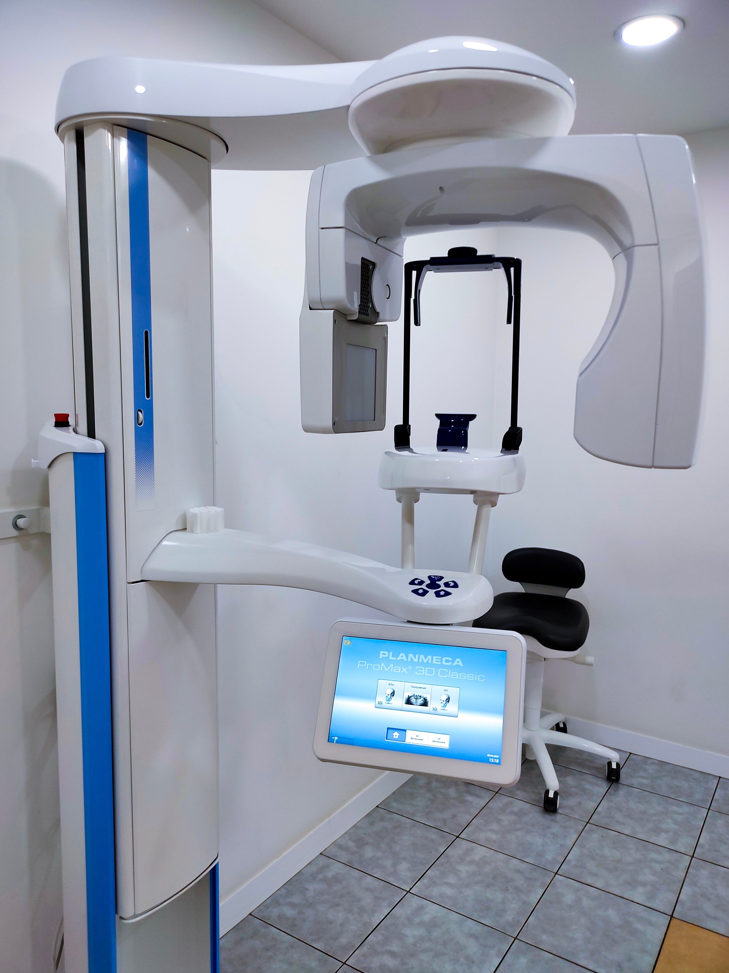
Cone Beam CT (CBCT)
Cone Beam CT, commonly known as CBCT, is a specialised form of dental imaging that produces detailed three-dimensional images of the teeth, jaws and surrounding facial structures.
It provides more detailed information than a standard dental X-ray and may be requested when your dentist, orthodontist or specialist needs to assess anatomy from multiple angles.
Available At
What does CBCT examine?
CBCT can provide detailed images of individual teeth and tooth roots, the upper and lower jaws, impacted teeth, bone surrounding the teeth, the jaw joints, parts of the sinuses and facial bones, and other structures relevant to dental treatment.
Why might I need a CBCT?
Your dentist or specialist may refer you for CBCT to assist with dental implant planning, assessment of impacted or unerupted teeth, root canal or endodontic assessment, orthodontic treatment planning, assessment of the jaw and facial bones, investigation of complex dental problems, or planning for oral or maxillofacial surgery.
What can I expect?
You will be positioned at the CBCT scanner with your head supported to help you remain still.
The scanner moves around your head while a series of images are taken. These images are then combined to produce detailed three-dimensional views of the area requested by your referring practitioner.
The examination is quick and painless.
How should I prepare?
No special preparation is usually required.
You will need to remove jewellery, glasses, removable dental appliances, hair clips and other metal objects around your head or neck where possible, as these can affect image quality.
Please tell our team if you are pregnant or think you may be pregnant.
Benefits of CBCT imaging
CBCT provides detailed three-dimensional information about dental and facial structures that may not be visible on conventional dental X-rays. This can help your dentist or specialist assess complex anatomy and plan treatment more precisely.
FAQ’s
-
Standard dental X-rays generally provide two-dimensional images, while CBCT produces detailed three-dimensional images that can be viewed from multiple angles.
-
CBCT and conventional CT both produce cross-sectional images, but CBCT is specifically designed for detailed imaging of dental and facial structures.
-
The actual scan is usually very quick, although additional time is required to position you correctly.
-
Yes. CBCT uses X-rays to create the images. The examination is performed using an imaging protocol appropriate to the area your practitioner has requested.
Dentascan
Dentascan is a specialised CT examination used to create detailed images of the upper or lower jaw.
It provides cross-sectional and reconstructed images of the jaw and surrounding structures and is commonly used when detailed information about the jawbone is required for dental treatment planning.
Available At
What does a Dentascan examine?
A Dentascan provides detailed images of the upper or lower jaw, jawbone, tooth roots and surrounding structures, the position of important anatomical structures, and areas being considered for dental implants or other procedures.
The images can be reconstructed in different views to help your dentist or specialist assess the height, width and shape of the jawbone.
Why might I need a Dentascan?
Your dentist, oral surgeon or specialist may refer you for a Dentascan to assess the jaw before dental implant placement, determine the amount and position of available bone, assess important structures within the jaw, investigate complex dental or jaw concerns, or assist with planning dental or oral surgery.
What can I expect?
You will lie on the CT scanning table while your head is positioned for the examination.
The table moves through the wide opening of the CT scanner while the images are taken. You will need to keep still for a short period to ensure clear images.
The scan itself is generally quick and painless.
How should I prepare?
There is usually no special preparation required for a Dentascan.
You may be asked to remove jewellery, removable dental appliances or other metal objects around your head and neck where possible.
Please let our team know if you are pregnant or think you may be pregnant.
Benefits of Dentascan imaging
Dentascan provides detailed information about the jawbone and surrounding structures and can assist your dentist or specialist when planning implants, surgery or other dental treatment. The reconstructed images allow the area to be assessed from several different views.
FAQ’s
-
Dentascan is commonly used to assess the jawbone and surrounding anatomy when planning dental implants or other dental and oral surgical procedures.
-
Both examinations can provide detailed cross-sectional dental images. CBCT uses a dedicated cone-beam dental scanner, while Dentascan uses CT imaging with specialised dental reconstructions. Your referring practitioner will request the examination most appropriate for the information they need.
-
Dental Dentascan examinations generally do not require intravenous contrast.
-
The scan itself is usually very quick, although your overall appointment will include time for positioning and preparation.